What is an MRI scan?
MRI is a routine diagnostic imaging exam that uses a large magnet, radio waves, and a computer to produce two- and three-dimensional images of the body's organs, tissues, and bones. Most MRI machines are a large, tube-shaped magnet that provide a strong magnetic field around your child. A radio frequency coil is placed over the body part that is to be imaged. The magnetic field, along with applied radio frequency waves, temporarily alters the alignment of hydrogen protons found in water molecules within the body. Computers construct the images based on the radio frequency signals emitted by the protons.
MRI is often preferred over other imaging techniques because it does not use ionizing radiation (x-rays). It's a way to better evaluate various parts of the body and certain diseases that may not be assessed adequately with other imaging technologies, and it's painless and safe.
An MRI is interpreted by a pediatric radiologist or pediatric neuroradiologist, and the results are reported to your child's physician.
MRI with anesthesia
Sometimes, MRIs need to be performed under general anesthesia. Movement will cause the MRI pictures to be blurred. Your child must lie still during the MRI scan. The use of anesthesia will cause your child to go to sleep and remain motionless and comfortable during the scan.
How should I prepare for an MRI?
If your child is receiving general anesthesia, you will get a call from an assessment nurse before your appointment to review your child’s health history.
Ask a member of your health care team how to prepare for the MRI if your child has any surgical metal implants (you will need to get MRI safety documents and bring them to the test).
For patients who are NOT receiving anesthesia, one parent/guardian can stay in the room during the MRI.
For patients who ARE receiving anesthesia, one parent/guardian may be able to stay in the room while the anesthesiologist gives your child the general anesthesia. After that, you must leave the room before the MRI starts.
If you are pregnant, you cannot be in the MRI room with your child.
Explain in easy-to-understand terms what this test is and why it is being done (“We’re going to take pictures of your knee to see why it hurts”). Explain that your child will have to lie very still (“like a statue”) during the test so the doctor can get clear pictures.
Tell your child that:
- MRI scans can take anywhere from 20 to 90 minutes. The MRI team will give you a more specific timeframe on the day of the scan.
- They will change out of their clothing and into a hospital gown for the scan.
- The MRI machine makes loud banging and clicking noises during the scan, but it will not hurt them.
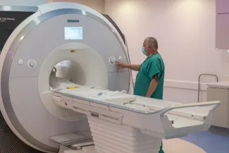
What happens during the MRI scan?
Your child will be positioned on the scanning bed. The inside of an MRI machine looks like a tunnel. It is necessary for the body part that will be scanned to be in the center of the scanner, so the technologist will move the scanner bed into the tunnel until it is appropriately positioned. Your child will have earplugs to protect their ears because the MRI machine makes loud pulsing or knocking sounds.
An MRI technologist will perform your child's scan and, if your child is receiving anesthesia, the anesthesia team will continue to monitor your child.
Your child may watch a movie or listen to music during the scan. Your child can choose movies or music from our collection. If you bring a movie, give it to an MRI team member.
Sometimes, patients receive a substance called gadolinium during the scan, which is needed to provide additional information about some parts of the body. Gadolinium is given through the IV.
MRI scans consist of several sequences of a few minutes duration each that cumulatively take anywhere from 20 to 90 minutes, depending on the information required by the radiologist and your physician. We will give you a more specific time frame before the scan begins.
What is an MRI arthrogram?
An MRI arthrogram is two-part procedure, involving fluoroscopy. First, a special type of x-ray technology, called fluoroscopy, is used to take pictures of the joint after a contrast material has been injected into it. This allows the radiologist to see the soft tissue structure of the joint. The joint is then examined with MRI, a routine diagnostic imaging procedure that uses a large magnet, radio waves, and a computer to produce two- and three- dimensional images of the body's organs, tissues, and bones.
An MRI arthrogram can be performed on the following joints:
- ankle
- elbow
- hip
- knee
- shoulder
- wrist
Your physician may request an MRI arthrogram when a problem with your child's joint cartilage is suspected. An MRI arthrogram may be more useful than an x-ray, because it shows the surface of soft tissues lining your child's joint as well as the bones.
MRI is done in conjunction with an arthrogram, because it can obtain specific diagnostic information not provided by the arthrogram.
What happens after the scan?
If your child did not receive anesthesia, your child can go home when the MRI scan is done.
If your child received anesthesia:
- We take your child to the MRI recovery room after the scan. A nurse gives your child fluids through the IV.
- Your child may eat juice or crackers after waking up.
- Your nurse lets you know when your child is ready to go home. The nurse also gives you information on whom to contact with questions or concerns.
- Your child may be sleepy. Help your child to stay steady and not fall while walking.
- Your child will use a wheelchair to get to your car. We strongly recommend you do not take public transportation.
- A recovery room nurse will call the next morning to make sure your child has fully recovered from the anesthesia.
How will I learn the results?
The radiologist reviews the images and sends a report to your referring doctor within two to three business days. Your child’s doctor shares the results with you.
If there is a finding on the scan that needs attention right away, we will contact your referring doctor as soon as possible to plan the next steps.